
Blunt cardiac injury (BCI) is caused by a direct blow to the heart. Such symptoms vary from clinically quiet transitory arrhythmias to lethal heart wall rupture. The lack of precise criteria and a gold standard for laboratory analysis makes it challenging to diagnose traumatic cardiac damage. Treatment is individualized based on the degree of the damage and might range from EKG screening to sternotomy with sophisticated surgical repair.
Blunt cardiac injury (BCI) spans a wide range of pathologies, from medically insignificant transitory arrhythmias to fatal heart wall rupture. The most prevalent kind is “cardiac contusion,” i.e., damage to the myocardium, which is still debatable. The lack of a clear description and an established gold standard for testing complicates the diagnosis of cardiac contusion. Arrhythmia, aberrant heart wall motion, perhaps escalating to cardiogenic shock, and rupture of valves, the septum, or a ventricular, atrial, or septal wall are crucial problems in acute cardiac injury.
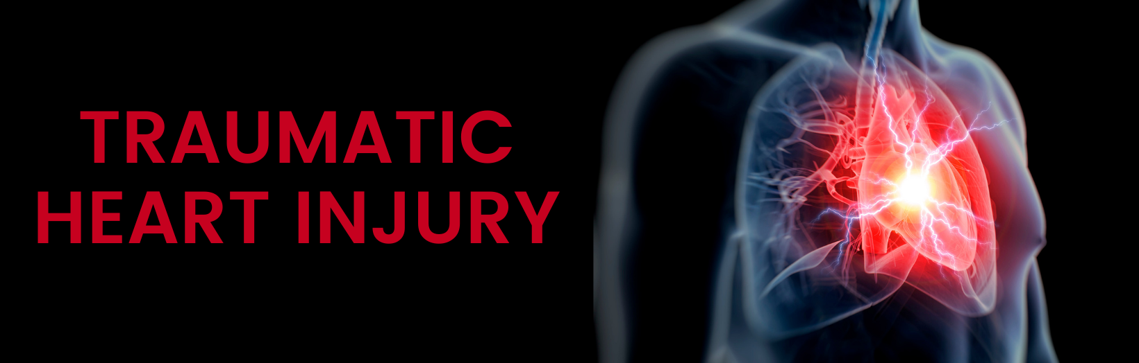
Blunt Cardiac Injury
Since the heart is securely encased within the bone thorax of ribs and sternum, tremendous power is required to induce BCI. This is particularly common in car accidents and situations of pedestrians being hit by cars. Other processes that contribute include falls, crush injuries, assault, and sports-related injuries, including direct impacts to the chest. Severe abdominal compression, on the other hand, might result in a quick surge in blood supply to the heart from the inferior vena cava, followed by a chamber burst due to an unexpected rise in intracardiac pressure.
Blunt coronary artery lesions are extremely rare. However, they can occur due to a direct hit, resulting in intimal disruption and thrombosis. This nearly invariably happens in association with a significant myocardial contusion, more commonly affecting the left anterior descending artery, which is located anteriorly in the chest behind the sternum. The consequences of such injuries can be disastrous, including myocardial infarction, emboli generation, arrhythmia, ventricular collapse, and prolonged ventricular rupture.
Valve injuries are also uncommon and are caused by blood compressive forces during contraction, resulting in valve, chordae tendonae, and papillary muscle rupture. The aortic valve is the most usually affected, followed by the mitral valve. These injuries are often characterized by a combination of left ventricular failure and cardiac arrhythmias.
Traumatic pericardial rupture is uncommon, yet it is the most serious type of blunt heart damage. It is caused by both direct contact to the chest and pressure changes from compressive force to the abdomen, resulting in pericardium laceration upon both diaphragmatic and pleural surfaces. This frequently happens parallel to the phrenic nerve and can result in herniation of the heart into the thoracic or abdominal cavities and torsing of the major arteries, leading to heart failure and fatality.
BCI is frequently accompanied by other thoracic injuries, like rib fracture, sternal fracture, pneumothorax, hemothorax, and pulmonary contusion, and is typically part of multisystem trauma. When these additional injuries are present, BCI should be expected, and these other injuries may significantly impact the patient’s individual prognosis.
Diagnosis
A strong index of suspicion, and a comprehensive examination of the cause, are required for the early detection of blunt cardiac damage. The vast majority of people have no symptoms. Those who do, complain most frequently report chest discomfort, which can be complicated by the existence of chest wall damage. More severe BCI may present as a shock, which must be separated from other types of hypotension like tension pneumothorax, neurogenic and hypovolemic shock.
CXR, EKG, Holter monitoring, cardiac enzymes, transthoracic and transesophageal (TEE) echocardiography, and nuclear medicine scans have all been used to diagnose BCI. Chest X-rays are regularly acquired in trauma patients and can reveal injuries to the rib cage, including rib fractures, frequently associated with BCI.
EKGs are a useful screening tool that can detect rhythm and conduction problems. There seems to be no pathognomonic discovery that can be used to diagnose BCI consistently.